
-
Physiology
Structure and Function4 Topics -
Lymphatics and Edema Formation
-
The Microcirculation
-
Vascular Control3 Topics
-
The Cardiac Cycle
-
Determinants of Myocardial Performance7 Topics
-
Neuro-Control of Heart and Vasculature4 Topics
-
Electro-Mechanical Association4 Topics
-
Electrical Side of the Heart4 Topics
-
PathophysiologyDefining Heart Failure
-
Causes of Heart Failure
-
MVO2 and Heart Failure
-
Cardiac Output and Heart Failure7 Topics
-
Compensation for Circulatory Failure
-
Vascular Tone in Heart Failure
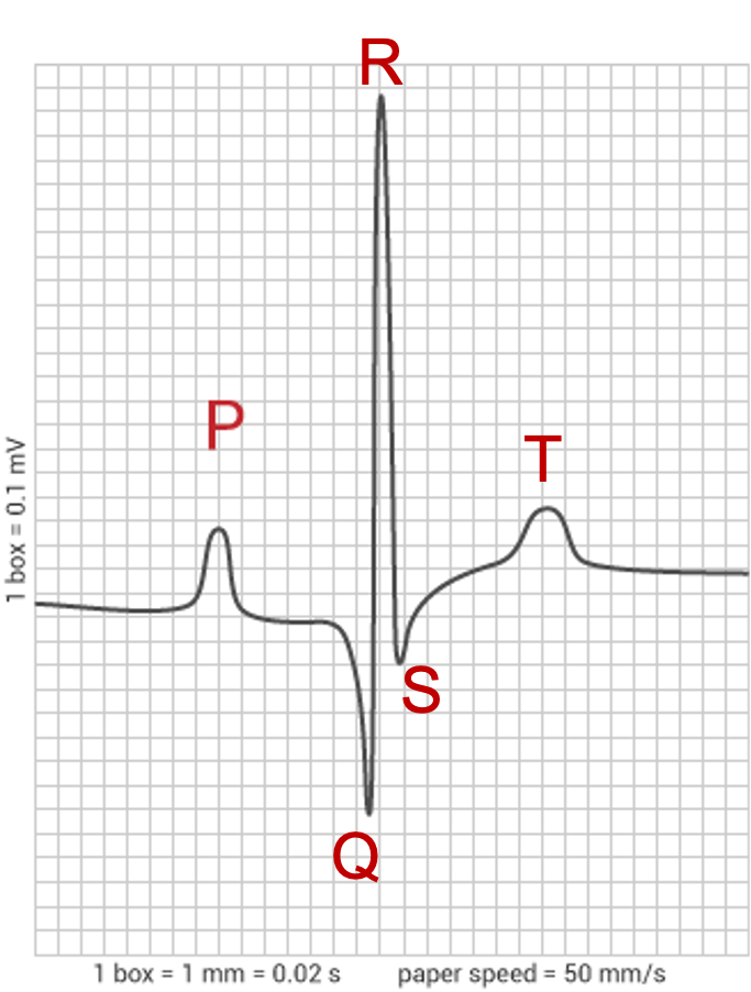
Just as the action potential is a graphic representation of the changes in membrane potential over time in a single myocardial cell, the electrocardiogram (ECG) is a graphic representation of the changes in potential in the whole heart over time. In the case of a normal rhythm, it is the product of a spontaneous depolarization originating in the SA node that conducts through the conduction system in a sequential and organized fashion, and resulting in a series of repeated waveforms. ECGs are most often recorded from the body surface.
Components of the normal ECG
P wave: Electrical activation (depolarization) of the atrial myocardium.
PR segment: This is a time of electrical quiescence during which the wave of electrical excitation (depolarization) passes through mainly the AV node. In addition the wave of depolarization moves through the bundle of His, bundle branches and Purkinje fibers. Since the wave of depolarization moves through the AV node at a speed of about 1/100th the speed the wave moves through the bundle of His, bundle branches and Purkinje fibers, most of the PR segment is associated with the passage of the wave of depolarization through the AV node.
PR interval: Represents the combination of P wave and PR segment activity.
QRS complex: Depolarization of the ventricular myocardium.
T wave: Repolarization of the ventricular myocardium.
Purpose of the ECG
The main purpose of the electrocardiogram is to identify cardiac rhythm abnormalities.
In animals with a Purkinje fiber distribution as represented by the dog, the ECG can also be used to make a statement about ventricular chamber enlargement.
Refer to the Electrocardiology section for more information on ECGs and interpretation.