
-
Physiology
Structure and Function4 Topics -
Lymphatics and Edema Formation
-
The Microcirculation
-
Vascular Control3 Topics
-
The Cardiac Cycle
-
Determinants of Myocardial Performance7 Topics
-
Neuro-Control of Heart and Vasculature4 Topics
-
Electro-Mechanical Association4 Topics
-
Electrical Side of the Heart4 Topics
-
PathophysiologyDefining Heart Failure
-
Causes of Heart Failure
-
MVO2 and Heart Failure
-
Cardiac Output and Heart Failure7 Topics
-
Compensation for Circulatory Failure
-
Vascular Tone in Heart Failure
Preload refers to the stretching of the myocardial cells in a chamber during diastole, prior to the onset of contraction. This is the priming process of the pump. Preload is measured as the end-diastolic volume or end-diastolic pressure.
Therefore, preload is equal to venous return plus the residual volume left in the cardiac chamber after the last contraction.
How do changes in preload change cardiac performance?
The Frank-Starling Law of the heart demonstrates the relationship between preload and stroke volume.
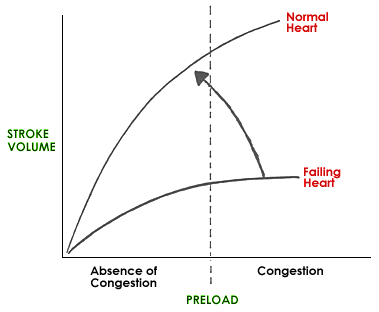
Frank Starling Curve 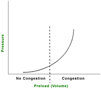
Pressure Volume Curve
The Frank Starling Curve shows that an increase in preload (end-diastolic volume) increases stroke volume. The Pressure-Volume Curve shows that as volume (preload) increases, pressure in that chamber increases. As the pressure in the chamber rises so too does the intravascular pressure (hydrostatic pressure) of the vessels that feed into this chamber. As preload increases and so pressure continues to rise, a point will be reached at which the ventricular end-diastolic pressure is sufficiently elevated to force the intravascular fluid in the vessels behind that chamber to leak out into the interstitium that is adjacent to these vessels. If this occurs in the lungs, pulmonary edema results. The vertical line in the figures refers to a hypothetical point, such that preload levels to the right of the vertical line promote the extravasation of fluid out of the capillaries into the interstitium (pulmonary edema if we are referring to left ventricular preload). Note also, however, that as preload falls (moving to the left along the curves) the forces that promote pulmonary edema are removed.
An increase in preload also increases afterload as the volume of the chamber increases.
The figure on the left shows two left ventricular function curves. Note how an increase in preload causes a marked increase in stroke volume for the normal heart but only a modest increase in stroke volume for the failing left ventricle. Conversely, reductions in preload cause a marked fall in stroke volume for the normal heart but only a modest reduction in stroke volume for the failing heart. Therefore, a marked reduction in preload in the heart failure setting will result in a resolution of pulmonary edema with only a modest reduction in stroke volume (but then these patients can’t afford a marked reduction in SV). Examples of therapeutic strategies used to reduce preload include diuretics and venodilators.
How are changes in preload detected on physical examination?
As left ventricular preload rises, pulmonary venous distention occurs and then pulmonary edema develops.
As right ventricular preload rises, central venous pressure increases causing jugular venous distention, hepatomegaly and ascites. In addition, pleural effusion may occur.
Excessive left ventricular preload may show
- pulmonary edema as detected by increased respiratory rate and effort, and pulmonary crackles (if severe) or increased broncho-vesicular sounds (if mild or moderate) on pulmonary auscultation
- hypoxemia/cyanosis
- signs of excessive right ventricular preload in addition
Excessive right ventricular preload may show
- jugular venous distention or distention of peripheral veins
- positive hepato-jugular reflux
- pleural effusion as detected by increased respiration rate and effort, and diminished lung sounds on auscultation
- hepatomegaly and/or splenomegaly
- ascites (abdominal fluid)
- subcutaneous edema