
-
Basics of ECG Interpretation11 Topics
-
Normal ECG Parameters
-
ECG Interpretation of Chamber Enlargement4 Topics
-
Dysrhythmias
-
Bradycardia
-
Heart Block3 Topics
-
Sick Sinus Syndrome
-
Tachycardia8 Topics
-
Hyperkalemia
-
Myocardial Hypoxia/Ischemia
-
Low Amplitude QRS Complex
-
Wide QRS Complex
-
Bundle Branch Block
-
Differentials for ECG Abnormalities
Lesson Progress
0% Complete
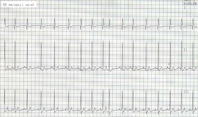
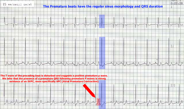
ECG Findings:
- Premature beats (beats that come earlier than expected)
- The morphology of these beats usually looks identical or very similar to the normal sinus beats
- The duration of the QRS complexes are usually normal
- They may or may not have an identifiable premature P wave associated with them
Etiology:
- These beats may originate within ectopic foci in the atria (atrial premature beats) or they may originate in the peri-AV nodal tissue
- The causes of both origins of supraventricular beats are identical:
- Due to disorders that result in enlargement of the atria (e.g. AV valve insufficiency)
- Due to inflammation, infection, ischemia, or neoplasia affecting the atria
- Drugs (digitalis, anesthetics)
- Extra-cardiac causes such as pain, other causes of catecholamine stimulation, intra-thoracic disease
Consequences:
- Several isolated premature beats are of no hemodynamic significance
- However, when enough of these beats are present, a reduction in cardiac output may result
Treatment:
- Several isolated beats do not warrant therapy. However, such individuals should be frequently monitored for exacerbation of their rhythm disturbance. In addition, attention should be directed toward identification and management of the underlying disorder.
- Criteria for therapy include:
- If the animal is symptomatic during the arrhythmia
- If paroxysms of sustained tachycardia develop such as a run lasting greater than 30 sec (see SVT below)
- Drugs used may include beta blockers (e.g. propranolol, metoprolol, atenolol), calcium channel blockers (diltiazem), sotalol, or digoxin.