Cardiovascular Physiology and Pathophysiology
-
Physiology
Structure and Function4 Topics -
Lymphatics and Edema Formation
-
The Microcirculation
-
Vascular Control3 Topics
-
The Cardiac Cycle
-
Determinants of Myocardial Performance7 Topics
-
Neuro-Control of Heart and Vasculature4 Topics
-
Electro-Mechanical Association4 Topics
-
Electrical Side of the Heart4 Topics
-
PathophysiologyDefining Heart Failure
-
Causes of Heart Failure
-
MVO2 and Heart Failure
-
Cardiac Output and Heart Failure7 Topics
-
Compensation for Circulatory Failure
-
Vascular Tone in Heart Failure
Distensibility
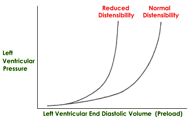
Distensibility refers to the ease of ventricular filling in diastole (ability to stretch). Lusitropy is the term used to refer to the distensible state of a cardiac chamber.
How do changes in distensibility change cardiac performance?
As a chamber becomes less distensible an equal end-diastolic volume (preload) causes higher chamber pressures. These are transmitted upstream and can lead to pulmonary edema if it is the left ventricle or left atrium that is affected with a reduced distensibility. Thus, for the same amount of preload the less distensible chamber generates higher pressures. Most cardiac disorders tend to reduce distensibility, some very much more than others. Chronic distention of a chamber, particularly the atria, results in an increase in distensibility.

How are changes in distensibility detected on physical examination?
Reduced distensibility of left ventricle or left atrium – may show evidence of left heart congestion:
- pulmonary edema causing increased respiratory rate and effort, crackles or increased broncho-vesicular lung sounds on auscultation
- hypoxia/cyanosis
- excessive right ventricular preload
Reduced distensibility of right ventricle or right atrium – may show evidence of right heart congestion:
- jugular venous distention or peripheral venous distention
- positive hepato-jugular reflux
- pleural effusion (increased or laboured respiration, diminished lung sounds)
- hepatomegaly and/or splenomegaly
- ascites (abdominal effusion)
- subcutaneous edema
Comments: Note how this looks like elevated preload in the adjacent upstream chamber. In fact, reduced distensibility causes a form of elevated preload in the adjacent upstream chamber.
