Improving Veterinary Radiology – Ending The Spray And Pray Mentality
We all know the feeling, a patient presents for a wellness check and your practice plan allows for radiographs, so the whole body is imaged. Or there is a left pelvic limb lameness so we imaged the femur, but then added in the spine for good measure. We think its a cruciate but we will include the whole abdomen with the stifle on the edge, just in case we catch something else. While this might seem like the best way to get a “bang for your buck” in diagnostic imaging, its actually a complete disaster. Improving veterinary radiology starts with a targeted approach.
When we image the whole dog, not only have we reduced the image quality, but we also end up with a lot of questionable findings. Put yourself in my position, I get a study which is a smorgasbord of anatomy and I need to sort through why we might have taken the images. Sure, the history says lameness, but was Fluffy also vomiting or does that stuff in the stomach not matter at all? I really want to give an answer. Radiologists don’t like to be fence sitters. I hate writing things like “unfortunately complete evaluation is precluded as the region of interest is not collimated in the available radiographs” or “probably a recent meal in the absence of a clinical history of gastrointestinal signs, however, foreign material cannot be excluded.”
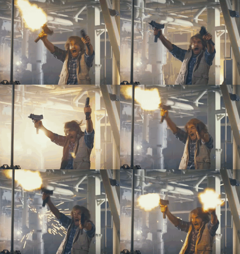
Essentially what I am pleading for is an end to the ‘spray and pray’ mentality of diagnostic imaging.

For anyone who didn’t come of age on a steady diet of Counterstrike at the local internet cafe, or is not a big Macgruber fan, ‘spray and pray’ is essentially what you see above. Just open the clip and hope you hit something.
If I see radiographs of the entire dog this is generally what I assumed happened. The animal presented with that classic “aint doing right” presenting signs and so we just went for it. I know its not always like that. I remember the panic of being in clinics without a radiologist. We just want that answer so we image a lot and hope we grab something. After all, we often do, and the radiologist can probably tell us something.
But what ends up happening is that we miss out on the important things, or we find things of unknown significance. A common scenario for me is to radiographs of a dog with a pelvic limb lameness. It is probably a cruciate, but because we tried to get the rest of the abdomen, the stifle superimposes that lovely para-preputial fat pad and we can’t see if there truly is effusion in the joint. And to top it off Lucky has multiple narrowed disc spaces. So are they clinically significant? Could it be IVDD causing the lameness?
Who knows?
In my opinion it ends up increasing cost to our patients if we need to reimage one of those findings. And it definitely increases the radiation exposure for your staff and for the patients.
So to improve veterinary radiology here is what I propose. Three well positioned views. That’s all we need.
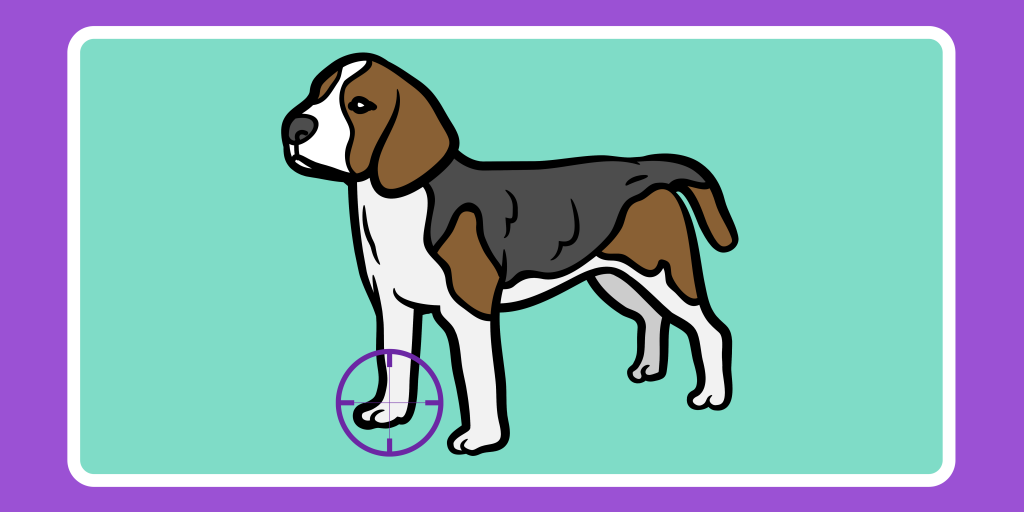
Start with your physical exam. When you find out where the problem might be take radiographs collimated to that anatomy.
Take both left and right laterals and a VD if its the thorax or abdomen.
This means that if its a limb we take a craniocaudal, a lateral, and then maybe one more (oblique, flexed lateral).
That’s it, no more, no less. The only thing we want is 3 well positioned views
If Rover is coughing and is lame, three views of the thorax and 3 separate views of the limb.
There are still going to be times when you need to image more of the body. But those times will be fewer and farther between. Collimate to your area of interest, get well positioned views and radiology is going to stop being that impossible to interpret thing you have to suffer through.
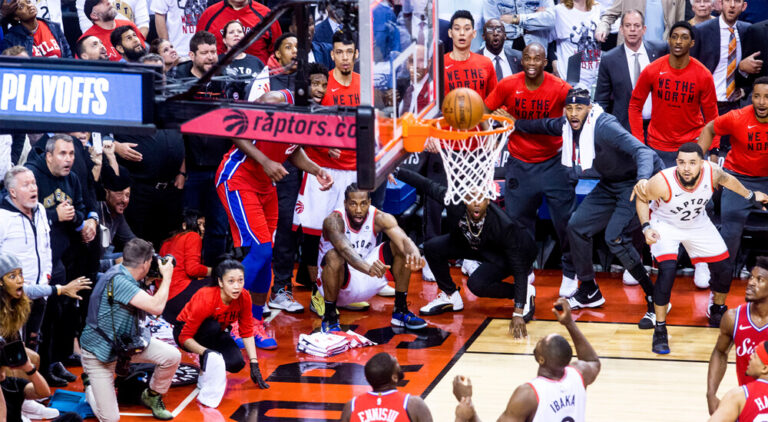
By taking these steps and improving veterinary radiology in your practice, you’ll feel less like Macgruber, and more like Kawhi Leonard from the corner. Deadly accurate and king of the world.


Responses