Lesson Progress
0% Complete

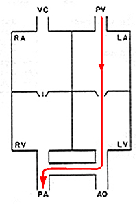
PDA Recirculation Circuit:
- The recirculation circuit is the path that a red blood cell would take if it continuously traversed across the defect on every occasion when it encounters the defect.
- The structures within the recirculation circuit are subject to volume overload; that is they carry an increase in blood volume.
- The cardiac structures within the recirculation circuit respond to volume overload by eccentric hypertrophy (dilation).
- The arteries and veins within the recirculation circuit respond to volume overload by dilation.
- For a left to right PDA the recirculation circuit is: the main pulmonary artery; the pulmonary arteries; the pulmonary veins; the left atrium; the left ventricle; the ascending aorta; the arch of the aorta; and the descending aorta up to the ductus arteriosus.
The abnormality:
- Failure of the ductus arteriosus to close shortly after birth and thereby allowing continued flow of blood between the aorta and pulmonary artery.
- Ductal smooth muscle hypoplasia is responsible for failure of the ductus to close.
- A polygenic mode of inheritance is proposed for Miniature and toy poodles.
The Incidence:
PDA is reported to be the most frequently encountered congenital cardiac defect in the dog. We now believe that aortic stenosis is the most common cardiac congenital disorder in the dog.
1. Classical Patent Ductus Arteriosus (PDA) (Left to Right Shunt):
- most cases of PDA involve blood flow from the higher pressure region (aorta) to the lower pressure region (pulmonary artery)
2. Reverse PDA (Right to Left Shunt):
- This may occur secondary to a hypoplastic pulmonary artery disorder or pulmonary artery hypertension resulting in a markedly elevated pulmonary artery pressure such that the pulmonary artery pressure is greater than the pressure in the aorta.
- In people, a reverse PDA may occur secondary to a large volume of blood flowing into the pulmonary artery due a left to right shunt. Chronic left to right shunting of blood is reported to increase pulmonary artery tone and result in elevated pulmonary artery pressure. To the authors’ knowledge this has never been demonstrated to occur in the dog.
