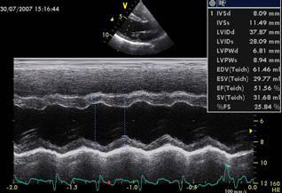
This is the oldest form of echocardiography. It was introduced into Veterinary Medicine in the late 1970s. It is more specifically known as the Time Motion Mode.
This view of the heart provides information about a very narrow core of tissue. The dimensions of this narrow core of cardiac tissue can be readily obtained over numerous cardiac beats in this format. Although this form of echocardiography was the first to be described, it is still best suited to measure the thickness of the ventricular walls and the dimensions of the internal cavity of the ventricles. From these parameters indices of the ventricular contractility can be readily determined. Of these indices, fractional shortening [FS] (left ventricular internal dimension in diastole [LVID-D] minus left ventricular internal dimension in systole [LVID-S] all divided by the left ventricular internal dimension in diastole [LVID-D] is the most common. The normal value for FS in the dog and cat is 25 to 45 %. Other indices of contractility are under consideration as to their utility, such as:
- the mitral valve “E” point to septal separation [EPSS],
- the ratio of the LVID-D to the left ventricular free wall dimension in diastole [LVFW-D],
- the ratio of the LVID-S to the left ventricular free wall dimension in systole [LVFW-S].
As well as these derived parameters there are numerous other indices that have been proposed to assess the performance of the heart. The dimensions of the internal diameter of the ascending aorta and the left atrium can also be determined. As well, M-mode Echocardiography is ideal to closely examine for abnormalities in the motion of the valve leaflets (such as fluttering of the anterior leaflet of the mitral valve in diastole associated with aortic valve insufficiency or premature closure of the aortic valve associated with left ventricular concentric hypertrophy) which may suggest a specific cardiac morphologic or physiologic abnormality.
Even though M-mode Echocardiography represents the oldest form of this imaging modality, M-mode Echocardiography will continue to have an important role to play in the assessment of cardiac disorders a result of its great ability to readily and repeatedly quantitate intracardiac linear dimensions.
It must be noted that M-mode Echocardiography requires considerable expertise to perform properly. The most technically demanding feature is to identify the exact positions in the heart that fulfill the criteria established to obtain the specific parameters to be measured. Furthermore, the specific location for calaper placement to properly measure the desired parameters has been rigidly established. For adequate interlaboratory comparison of data, the established criteria must be rigidly adhered to.
