
This is the newest form of echocardiography. Doppler Echocardiography is an additional and supplemental part of an echocardiographic examination. Doppler involves the detection of blood flow. During an echocardiographic examination, the Doppler examination detects blood flow throughout the heart.
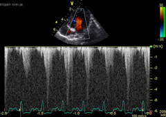
The location of normal and abnormal blood flow as well as the velocity of blood flow can be readily detected. The velocity of blood flow is displayed as a histogram of peak velocities over time.
This procedure is a completely non-invasive diagnostic test in all its various modalities. In the vast majority of cases even sedation is not required as an aid to perform this examination.
Assisting with the identification of cardiac disease
The Doppler Echocardiographic examination enables the specific identification of valvular heart disease; ie, valvular insufficiency and valvular stenosis. As well, the Doppler examination enables the detection of intracardiac shunts; ie, atrial septal defects and ventricular septal defects.
The Doppler examination also provides evidence of abnormal diastolic function, and reductions in cardiac output.
Differences from routine echocardiographic examination?
Although a Doppler examination can be performed independent from a two-dimensional examination, most Doppler echocardiography is performed simultaneously with a two-dimensional examination.
A routine two-dimensional echocardiographic examination provides spacial information about cardiac chamber size and wall thickness. Thus, with a two-dimensional examination the size of the left and right ventricle in systole and diastole can be measured, particularly with the aid of M-mode echocardiography, the size of the left atrium can be measured, and indices of contractility can be derived. And so we tend to infer the existence of valvular disease based on changes in the volume of the chambers or the thickness of the walls. Sometimes two-dimensional echocardiography may reveal the existence of reduced valve leaflet excursion as in stenotic valvular disease, or prolapse of valve leaflets as in valvular insufficiency. However, as opposed to such indirect evidence, Doppler echocardiography provides direct evidence for the existence of valvular disease.
Insight into the severity of valvular heart disease
One of the greatest assets of Doppler echocardiography is its ability to assess the severity of the valvular disorder. In the case of stenotic valvular disease, as the stenosis progresses the peak velocity of blood flow across the stenotic lesion increases. Thus stenotic disorders can be readily classified as to severity with the determination of the Doppler velocity of blood flow.
The normal peak velocity of blood flow across the four valves in the heart has been determined for the dog:
- Tricuspid valve = 0.9 meters/sec
- Mitral valve = 1.1 meters/sec
- Pulmonic valve = 1.2 meters/sec
- Aortic valve = 1.5 meters/sec
The ability of Doppler echocardiography to quantitate the severity of valvular insufficiency has been more ambiguous. It would appear that Doppler echocardiography may only be useful to provide a semi-quantitative assessment of the severity of valvular insufficiency.
Detection of atrio-ventricular valvular insufficiency
Doppler echocardiography employed to detect atrio-ventricular valvular insufficiency is primarily performed from the left parasternal position. In the Doppler (spectral display graphic display of the peak velocity histograms), the turbulent signal is negative and occurs in systole.
A lesion is classified as mild if the insufficiency signal is detected only in the region close to the atrio-ventricular valves. A lesion is considered to be of moderate severity if the signal is detected into the mid-region of the affected atrium. And finally, the insufficiency is classified as severe if the turbulent signal of atrio-ventricular valve insufficiency is detected at the base of the affected atrium or in the inlet vessels to the affected atrium.
The incidence of tricuspid valve insufficiency, as determined by Doppler echocardiography, has been reported to be approximately 50% in normal dogs.
Detection of semilunar valve insufficiency
The pulmonic valve may be interrogated from the right and left parasternal positions with Doppler echocardiography. The aortic valve is examined from the left parasternal position with Doppler. In the Doppler spectral display, the turbulent signal of semilunar valvular insufficiency occurs as a positive signal and in diastole.
Mild lesions result in the detection of turbulence only in the region of the outflow tract proximal to the semilunar valves. With moderate to severe lesions the turbulent signal is detected deep into the chamber of the ventricle.
Pulmonic valve insufficiency as detected by Doppler echocardiography occurs in 25 to 70% of normal dogs.
Detection of atrio-ventricular valve stenosis
The Doppler examination is performed from the left parasternal position. The Doppler spectral display reveals an increased peak velocity that is positive and occurs in diastole.
Detection of semilunar valve stenosis
The pulmonic valve may be examined from the left or right parasternal position. The aortic valve is examined from the left parasternal position. Stenosis is present when the peak velocity of blood flow detected across the outflow tract is increased. The Doppler spectral display reveals a negative, systolic signal that is increased.
As the severity of the stenosis progresses, the peak velocity of blood flow across the stenosis increases.
Presence of diastolic disease of the ventricle
Diastolic disease of the ventricle is commonly one of the earliest signs of heart disease. Diastolic disease of the left ventricle is demonstrated by the presence of left atrial enlargement and later pulmonary edema. Diastolic disease of the right ventricle is indicated by the presence of right atrial enlargement and later jugular distention or ascites.
The presence of increased velocity of blood flow across the atrio-ventricular valve that normally occurs in late diastole and is associated with atrial contraction suggests the presence of reduced ventricular compliance (the Doppler E wave wave).
Additional disorders detection with Doppler Echocardiography
A ventricular septal defect can be detected with Doppler echocardiography. With a left to right shunt, the peak velocity of blood flow is inversely related to the severity of the defect.
Left to right patent ductus arteriosus can be diagnosed by detecting a continuous turbulent signal in the main pulmonary artery.
Cases of reduced cardiac output are associated with reduced velocities of blood flow across the aortic valve.