
Gallops
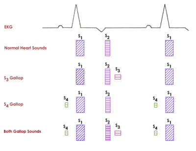
Gallops are low frequency diastolic sounds that result in a tripling or quadrupling of the heart sounds, resembling the canter or gallop of a horse. They are best heard with the bell of the stethoscope.
While the term ‘gallop rhythm’ is often used, it is a misnomer since a gallop is not a disorder of cardiac rhythm or an arrhythmia. Rather they are extra sounds associated with abnormal heart filling, and not electrical events or extra heart beats.
S3 gallop
- Low frequency sound
- Occurs shortly after the S2 sound, at the beginning of diastole during the rapid filling phase (after the T wave on ECG)
- Also referred to as a ventricular gallop
- Not normal in dogs and cats
- Indicates diastolic dysfunction
- Associated with reduced compliance of the ventricle while filling under conditions of high filling pressures, often in conditions of diastolic volume overload (e.g. dilated cardiomyopathy (DCM), severe valvular insufficiency)
- Caused by the sudden termination of longitudinal expansion of the ventricular wall during brisk early diastolic filling during the period of rapid ventricular filling
- Indicates significant cardiac disease
S4 gallop
- Low frequency sound
- Occurs shortly before the S1 sound, at the end of diastole during atrial contraction (after the P wave on ECG)
- Also referred to as an atrial gallop
- Not normal in dogs and cats
- Indicates diastolic dysfunction
- Associated with the atria trying to force blood into an already over-distended ventricle or because the atria are forcing blood into a stiff ventricle. Atrial contraction is required for an audible S4 sound. Thus it cannot occur in atrial fibrillation
- Occurs in disorders with impaired relaxation of the ventricle, typical of disorders characterized by concentric hypertrophy like hypertrophic cardiomyopathy (HCM)
- More rarely heard in older stressed cats without detectable cardiac disease
Clicks
Clicks are very high frequency abnormal sounds most often associated with valve leaflet or valve apparatus motion in particular abnormal underlying conditions. While they are uncommon overall in small animals, they are more commonly heard in dogs than cats.
When present in mid to late systole, they are most commonly associated with degenerative mitral valve disease, and specifically likely prolapse of the mitral valve (cupping of mitral valve leaflet backwards into the left atrium in systole).
When present in early systole, they may be associated with aortic or pulmonary valve opening in aortic or pulmonary stenosis.
Split Heart Sounds
Normally the two AV valves close synchronously resulting in a single S1 sound, and the two semilunar valves close synchronously resulting in a single S2 sound. This is a result of the right and left ventricles contracting and ejecting simultaneously. Under conditions producing asynchronous closure of these valves (closure at slightly different times), either S1 or S2 may become split into two distinct sounds.
- A split S1 is due to asynchronous closure of the AV valves, which may occur with electrical conduction disturbances such as bundle branch block or with ectopic ventricular beats (both cause asynchronous ventricular contraction), or with mechanical disturbances in valve closure such as mitral or tricuspid stenosis.
- A split S2 is due to asynchronous closure of the semilunar valves. This may rarely be heard in normal large breed dogs during inspiration as there is more right ventricular filling and longer right ventricular ejection time during inspiration. Abnormal causes of split S2 include:
- conduction disturbances such as bundle branch block and ectopic ventricular beats,
- delayed closure of the pulmonary valve such as with pulmonary hypertension, pulmonary stenosis, or an atrial septal defect, or
- delayed closure of the aortic valve such as with systemic hypertension or aortic stenosis.
Friction Rubs
Friction rubs are abnormal extra sounds generated by rubbing of abnormal, often inflamed, pleural or pericardial surfaces against one another. They are very rare in small animals and more common in large animals, likely because of the nature of the underlying diseases that produce these sounds (e.g. pleuritis and pericarditis).
Friction rubs are described as scratchy, grating, or creaky sounds. They may be short or long in duration, and may have periodicity with the heart rhythm (pericardial friction rub) or with respiration (pleural friction rub).
They are caused by fibrin present on the pleural surfaces of chest wall, lungs, or pericardium.
Arrhythmias
The sympathetic stimulation produced by stress or anxiety results in many normal dogs and most normal cats having a regular rhythm on auscultation in hospital. Calmer patients with less sympathetic drive allowing more of the normal resting parasympathetic tone may have a normal sinus arrhythmia, characterized by phasic speeding up and slowing down of the heart rate, often coincident with respiration (speeding up on inspiration and slowing down on expiration). This is a normal finding and much more commonly appreciated in dogs than in cats, since dogs are more often calm in the hospital setting than cats.
Abnormal arrhythmias are the product of electrical abnormalities in cardiac rate and/or rhythm, and may produce transient or more sustained disruptions in rate and/or rhythm. Those that are associated with transient or sustained increases in heart rate above the normal sinus rhythm are called tachyarrhythmias, while those associated with a heart rate that is lower than the normal sinus rhythm are called bradyarrhythmias. Some forms of arrhythmia may be composed of both fast and slow components.
Tachyarrhythmias
- Isolated premature beats or pairs or short bursts of premature beats result in transient disturbances to the cadence of the heart rhythm.
- Sometimes a premature beat may result in only the presence of abnormally long pause between beats. This is because if the premature beat is extremely early (premature), no additional heart sounds may be detected, and an abnormal pause results. One might suspect that so-called “dropped beats” are occurring. Premature ventricular beats (PVCs) tend to cause longer pauses than supraventricular ones (SVPCs), but it is important to recognize that the type of premature beat (ventricular vs supraventricular) cannot be determined from listening – an ECG is required for this.
- If the premature beat is slightly less premature, only an S2 may be detected, so that the rhythm will sound like S1 then S2 and another S2 in rapid succession. This could be misinterpreted as a gallop. Gallops are not intermittent like premature beats and they are not followed by an abnormal pause.
- Even less premature beats may be associated with both an early S1 and S2.
- Sustained ventricular or supraventricular tachycardia (many premature beats in a row) can be associated with a regular rhythm, albeit at a faster than normal rate.
- Atrial fibrillation produces a very irregular rhythm and typically a fast heart rate (often described as sounding like “shoes in a dryer”). It may be difficult to differentiate from very frequent premature beats on auscultation and an ECG is required for definitive diagnosis.
Bradyarrhythmias
- Some bradyarrhythmias produce a heart rate/rhythm that is both slow and irregular. Examples can include some forms of atrioventricular block, some cases of atrial standstill, some cases of sinus bradycardia, and sick sinus syndrome (characterized predominantly by abrupt and lengthy pauses).
- Other bradyarrhythmias may produce a heart rate that is slow but a rhythm that is still quite regular. Examples can include some forms of atrioventricular block, some cases of atrial standstill, and even some cases of sinus bradycardia.
Abnormal Respiratory Sounds
Normal breath sounds are typically very quiet and sometimes barely heard particularly on expiration, depending on breed conformation and body condition. They should be soft, smooth, low-pitched, blowing sounds.
Abnormal respiratory sounds may arise from anywhere in the upper or lower respiratory tract. Types of abnormal respiratory sounds include the following:
- Stertor is a low-pitched snoring noise arising from the pharynx (often from flaccid tissue vibrating), audible without a stethoscope
- Stridor is a higher-pitched noise arising from the larynx or trachea (from rigid tissue vibrating), again often audible without a stethoscope
- Both stertor and stridor are upper respiratory noises associated with partial obstruction of the upper airways and not associated with cardiac disease.
- Wheezes are high-pitched continuous (long) inspiratory or expiratory sounds (but more commonly expiratory) that occur with tracheal or bronchial narrowing (either anatomic/structural or due to secretions)
- Crackles (formerly called rales) are high-pitched discontinuous (short) popping inspiratory sounds typically detected at the end of inspiration
- Subtle crackles may only be detected with a deep inspiration
- Sound like Velcro pulled apart, cellophane crinkling, or radio static
- Caused by explosive opening of small airways
- May be due to pulmonary edema (cardiogenic or non-cardiogenic), small airway disease (bronchitis), pulmonary fibrosis, or pneumonia, among other disease processes
- Crackles do not automatically indicate edema fluid in the the airways and do not equate to congestive heart failure. In fact, the loudest crackles are often associated with primary airway disease as opposed to pulmonary edema from heart disease.
- Diminished or absent respiratory sounds may also be considered abnormal, particularly when not explicable by increased body condition or when accompanied by increased effort. Pleural effusion is the most common cause, though other causes of sound insulation like hernias or mass effects may be considered.