Radiography is a simple and effective means of diagnosing cardiac chamber enlargement. In most forms of heart failure cardiac enlargement is present.
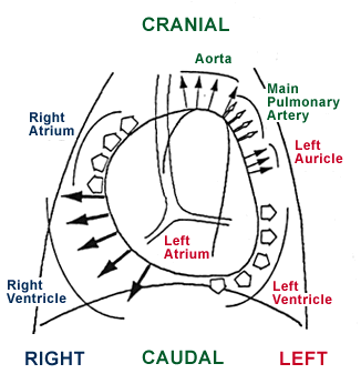
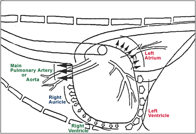
It is important to determine the cardiac structures that contribute to the silhouette of the heart on the lateral and D/V or V/D view.


Always be consistent with your lateral (R or L) and D/V or V/D views. Always obtain films at end inspiration.
Some general guidelines on cardiac size in the dog (criteria from end inspiratory films):
On the lateral view normal cardiac dimensions:
- horizontal plane:
- < 3.5 intercostal spaces for:
- brachiocephalic breeds
- immature dogs
- small breeds
- < 3 intercostal spaces for “average dog”
- < 2.5 intercostal spaces for deep chested breeds
- < 3.5 intercostal spaces for:
- vertical plane:
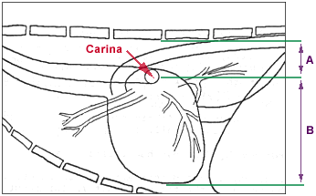
- the vertical distance from the cardiac apex to the carina is normally 2/3 to 3/4 the vertical distance from the cardiac apex to the vertebral column
- as cardiac enlargement occurs in this plane the trachea tends to parallel the vertebral column and the vertical distance from the cardiac apex to the carina is increased

A schematic diagram of a lateral radiographic view of the chest. Normally A is approximately 1/3 to 1/4 of A + B, and B is approximately 2/3 to 3/4 of A + B.
On the D/V or V/D view normal cardiac dimensions:
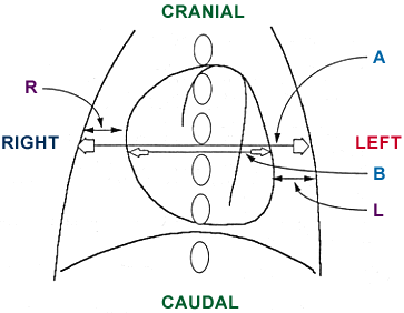
- the greatest horizontal cardiac dimension should be < 2/3 of the chest wall to chest wall thoracic dimension at that location.

A schematic diagram of a V/D or D/V radiographic view of the chest. In normal hearts R is approximately equal to L; B is < 2/3 of A.
Significance of cardiac enlargement in the lateral view:
- In the Horizontal Plane:
- Suggests right ventricular enlargement, however left ventricular enlargement tends to produce at least mild enlargement in this plane.
- In the Vertical Plane:
- Suggests left ventricular enlargement, however right ventricular enlargement tends to produce at least mild enlargement in this plane.
Significance of cardiac enlargement in the V/D or D/V view:
- A reduced right heart to chest wall dimension suggests right ventricular enlargement.
- A reduced left heart to chest wall dimension suggests left ventricular enlargement.
Single chamber enlargement is unusual and therefore enlargement in one chamber tends to cause enlargement in other areas of the heart.
Some general guidelines on cardiac size in the cat:
- The heart is more elongated and elliptical in shape than in the dog on the lateral view.
- The ventricular area occupies about 2 to 2 1/2 intercostal spaces in the lateral view.
- In the lateral view the cat heart tends to be more horizontal; as cats age, the heart tends to horizontalize even more (called a “lazy” heart).
- In the D/V or V/D projection, the cat heart is more oval and thinner than the dog. The cardiac apex usually lies on the midline.
