
The normal heart sounds are short, crisp sounds associated with vibration of cardiac structures during normal cardiac filling and emptying. In small animals, each heart beat is composed of two normal heart sounds, S1 and S2.
Let’s examine normal heart sounds a little more closely in terms of timing and qualities.
A phonocardiogram is a graphic recording of heart sounds, with time on the x-axis and sound amplitude (intensity) on the y-axis. It can be recorded with a simultaneous electrocardiogram (ECG), which is very helpful for appreciating the timing of the heart sounds relative to the electrical events, and in turn the mechanical events.
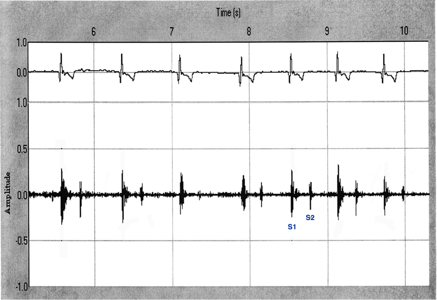
S1
- Associated timing-wise with closure of the atrioventricular (AV) valves (mitral and tricuspid) at the onset of systole and coinciding with the QRS complex on the ECG
- High frequency sound, typically louder than S2
- Heard best over the left apex
S2
- Associated timing-wise with closure of the semilunar valves (aortic and pulmonary) at the end of systole and coinciding with the end of the T wave on the ECG
- High frequency sound, typically sharp and higher-pitched
- Heard best over the left base
Interpreting the heart sounds in light of the cardiac cycle and the normal electrical and mechanical events is the first step to being able to appreciate and understand deviations from normal. Bringing it all together, S1 marks closure of the AV valves and the onset of systole, the ejection phase. The ventricles pump blood out into the great vessels during systole, producing the arterial pulse. So the arterial pulse is palpable after S1 (between S1 and S2). S2 marks the closure of the semilunar valves and the end of mechanical systole. The period between S2 and the next S1 is diastole, the phase when the heart fills.