Lesson Progress
0% Complete
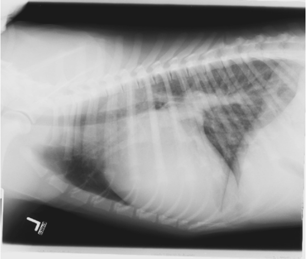
As the left side of the heart fails, that is less blood is ejected and venous return continues, then preload (fluid volume) to the left side of the heart becomes elevated. The elevated volume in the pulmonary venous system results in dilation or pulmonary venous engorgement. As the volume continues to increase, pressures in the pulmonary venous system become elevated and also in the pulmonary capillary bed resulting in a net efflux of fluid into the pulmonary interstitium. Venous engorgement is the first sign of increased volume in the left heart, making their detection on radiographs extremely important.
Radiographic signs of pulmonary venous distension:
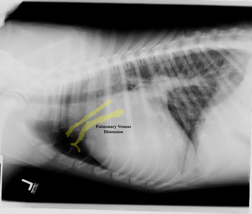
- In the lateral view, the cranial pulmonary veins are greater than the width of the the fourth rib
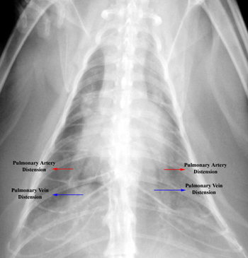
- The pulmonary vein is obviously larger than its accompanying pulmonary artery (normally they are of equal width)