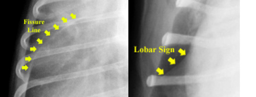
Both fissure lines and lobar signs appear as lines along the lobes of the lung, they both occur at the same location (along a lung lobe fissure) but they are easily differentiated. Radiographically, a fissure line is observed as a single white line due to the wicking of fluid between the two lung lobes (image: left below). A lobar sign is represented by a white margin adjacent to an area of radiolucency. This white margin represents the edge of a lung lobe that is consolidated often due to the presence of exudates, edema and/or hemorrhage. These are important to differentiate because they represent different disease processes. One represents fluid collecting in the pleural space (fissure line due to fluid) and the other represents flooding of the alveoli with fluid (note that exudates and hemorrhage are essentially fluid).
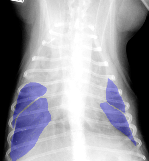
Increasing volumes of effusion widen pleural fissures creating more soft tissue opacity in the pleural space. This can cause retraction of lung lobes as noted below.