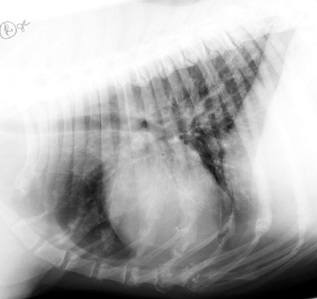
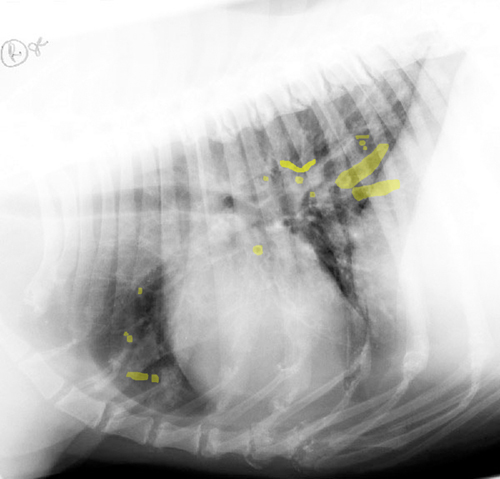
As mentioned previously the elevated capillary hydrostatic pressure causes fluid to “leak” into the pulmonary interstitium where it collects along the triad of artery, bronchus, and vein causing the so-called “bronchial pattern”, and silhouetting of the margins of the pulmonary arteries and veins. With respect to the development of a bronchial pattern, the fluid weeps down both the inside and outside of an airway. The walls of the tertiary bronchioles are not detectable radiographically due to the fact that they no longer have cartilaginous rings. However, once the inner and outer wall of the bronchiole is covered in fluid, it becomes more radiopaque and therefore contrasts very well against the radiolucent lumen (filled with air). As a result, on cross section the bronchiole appears as a radiopaque circle with a radiolucent center (“donut”), and if cut longitudinally it appears as two parallel radiopaque lines (“railroad track”).
Bronchial patterns are not specific for airway disease and can reflect primary causes of bronchial thickening